Library search
Search Results
-
 . A number of studies in recent years highlight the evidence to support the hypothesis that patients with chronic cystitis of different etiology are predisposed to bladder cancer (BCa) development. At the same time, today the problem of the influence of chronic urinary tract infections (UTI) on the clinical course of BCa is not well understood. Objective. The aim of our study was to investigate the effect of UTI on the clinical course of transitional cell carcinoma of the bladder. Materials and methods. The study was conducted on the basis of a retrospective analysis of case histories of 366 patients with bladder tumors admitted to the urological department for surgery. The Kaplan-Meier method was used to compare relapse free survival, time to tumor progression, downgrade and final treatment option (radical or palliative) in BCa patients with or without concomitant UTI. The log-rank test was used to assess the statistical significance between the two groups. Cox regression analysis with calculation hazard ratio (HR) 95% confidence intervals (CI) was used to predict the risks of above mentioned end points in comparing groups. Results. Symptoms of urinary tract infections were detected in 144 (39.3%) patients with bladder tumors, among them - in 124 (41,3%) with bladder cancer. Univariable and multivariable analysis showed a statistically significant increase in the risk of BCa relapse in the presence of concomitant UTI (HR = 1.88 (95% CI 1,24-2,85), p = 0,003 and HR = 1.91 (95 % Cl 1,24-2,93), p = 0,003, respectively. Median survival time without relapse was 1 422 and 2 120 days for the group with concomitant UTI and without it, respectively,. Thus, a longer dis ease-free survival was observed in patients without concomitant UTI (p = 0,002). No statistically significant differences in survival before the progression, downgrade and final treatment, depending on the presence of UTI were found (p = 0, 263, p = 0,177, p = 0,503, respectively). Conclusions. 1. Chronic urinary tract infection occurs more than in one-third (41,3%) patients with bladder cancer. 2. The presence of concomitant urinary tract infections in bladder cancer patients is an independent risk factor for recurren ce of the tumor. 3. There were no convincing evidence to support the effect of urinary tract infections in the progression, reducing the level of differentiation and reduction in time to carry out radical treatment
 -

The lack of understanding of bilateral breast cancer is a much more complex problem for oncologists. Today the incidence of unilateral as well as bilateral breast cancer is much higher than 10 years ago, which makes this problem even more urgent. Information about the causes of bilateral breast cancer, important criteria for the development of the disease, early diagnosis of the disease and early detection of the possibility of bilateral metachronous cancer, as well as preventive measures are not fully covered in the literature, scientific studies. In this article, the authors analyze the data of the recommendations of the international scientific society and the results of large clinical trials on bilateral breast cancer. Also, important criteria in the development of metachronous and synchronous breast cancer were analyzed, the results of clinical and morphological, immunohistochemical aspects were studied. The reasons for the development and modern knowledge about the diagnosis of bilateral breast cancer have been studied. In particular, the authors reviewed and studied about 40 foreign and domestic scientific works devoted to this problem.
-
Epidemiology, Risk Factors, Clinical-Imaging Features And Priorities For The Prevention Of Esophageal Cancer In The Fergana Valley Of Uzbekistan
The American Journal of Medical Sciences and Pharmaceutical ResearchCancer of the digestive system is the most common cause of death among malignant neoplasms (Table 1). According to the International Agency for Research on Cancer (IARC) for 2008, the incidence of cancer of the digestive system was 49.2 people per 100 thousand people per year, the mortality rate was 34.3 people per 100 thousand. At a relatively low incidence rate, esophageal cancer is the seventh most common cause of death from malignant tumors, giving way to lung, breast, stomach, liver, prostate, and colon cancers. This is due to the extremely malignant nature of the course, early metastasis, and late diagnosis of esophageal cancer. The aggressiveness index, calculated as the ratio of deaths to new cases, is extremely high in esophageal cancer and is about 95%.
The absolute number of deaths from esophageal cancer in 2008 in the world was 406 thousand people. In developing countries, morbidity and mortality from esophageal cancer are significantly higher than in developed countries (Table 2). The most common two histological types of esophageal cancer are squamous cell carcinoma and esophageal adenocarcinoma. Despite the similarity of the clinical picture, diagnostic and therapeutic tactics, an extremely unfavorable prognosis for both forms of esophageal cancer, these malignant neoplasms have different risk factors, socio-geographic and ethnic characteristics, knowledge of which is necessary for the timely establishment of the diagnosis and preventive measures. Squamous cell carcinoma of the esophagus (Fig. 1) is an extremely aggressive epithelial malignant tumor of stratified squamous epithelium, in most cases localized between the middle and lower third of the esophagus, the tumor is rare in the cervical esophagus.
-
 Bladder cancer occupies leading place in the structure of oncourology in last years. It is increasing incidence of bladder cancer and year by year this pathology is more exists in young patients. In this manuscript authors presents their own experience: bladder cancer in young patients runs more favorably and in young patients the number of superficial tumors and histological high differentiated (G3) cancer meets more than in elderly age patients. Despite of age and differentiation degree it is necessary to approach with mind at the choice of operations.
 -
 Pathogenesis and transition of normal urothelium of bladder into cancer arc multifactorial processes. Chronic inflammation causes the initiation and progression of the main pathophysiology of invasive and metastatic cancer. Л dichotomy is observed in the role of immune cells in bladder cancer. In this review, we discussed genetic and immunological changes of bladder cancer along with relapse of this disease; however, it should be noted that in most cases, multiple genetic and immunological changes occur simultaneously or mutually dependent on each other. Many genetic mutations disrupt the function of genes involved in regulation, and conversely, chromosomal aberrations lead to changes in transcription. Although the immune response protects the bladder by suppressing tumor growth, certain immune cells including neutrophils, macrophages, and T-lymphocytes contribute the development and progression of the tumor.
 -
 Bladder cancer on the background chronic inflammation: immunohistochemical assessmentEffect of chronic inflammation caused by uropathogenic strains of bacteria for a bladder cancer is a little-studied subject. Objective was to reveal the presence of relations between expression of CD95, p53, Ki-67, Bcl-2, BAX, E-cadherin and β-catenin COX-2, eNOS и iNOS markers and clinicopathological characteristics of superficial bladder cancer in presence of concomitant urinary tract infection. Materials and methods. Immunohistochemical study of expression mentioned above markers in bioptates taken from 44 superficial bladder cancer patients who were divided into groups according to absence or presence of concomitant urinary tract infection. Results and conclusions. 1. Statistically significant increase of markers p53, Ki67 and decrease СD95, BAX, E-cadherin and β-catenin expression in tumor tissue as against the control was found. That testifies to decrease apoptotic activity and adhesive properties, increase of proliferation of urothelium. 2. Increased expression of COX-2 and iNOS in the tumor tissue creates conditions favorable for the further development of the tumor. Statistically significant increase in the expression of iNOS on the background of the inflammatory process may be evidence in favor of the negative effects of inflammation on the course of cancer. 3. Presence of strong positive relationship between p53 and Ki67, bcl-2 and BAX expression in I group; bcl-2 and Ki67, BAX, β- catenin expression in II group can testify about changing of the investigated factors' interaction and be the reason of tumoral development against the background of inflammatory process.
Journal problems of biology and medicine -
Some Shocking Stories: The Behavioral Response Of Cancer Mortality To Spikes In Alcohol And Tobacco Consumption
The American Journal of Applied sciencesTobacco and alcohol have long been acknowledged as carcinogens holding a critical role in the progression of a various cancers. Identifying the degree to which tobacco and alcohol can impact cancer mortality is necessary to developing effective public health strategies and mitigating risks with preventative measures. The goal of this study is to characterize the behavior of cancer mortality in response to shocks in alcohol and tobacco consumption utilizing aggregate U.S. data. This is the first study of its kind to examine the intertemporal relationship between cancer mortality and its determining factors within a dynamic system. Our results indicate cancer mortality displays persistence and its path dependency varies considerably between the shock factors. An unexpected shock to alcohol consumption results in cancer mortality taking about 17 years to return to its pre-shock level, whereas tobacco consumption shocks recover the original cancer mortality level in about 10 years. Alcohol has a more dominant effect on cancer mortality regardless of time dimension. As a result, policies that have been previously emphasized toward mitigating tobacco consumption may prove prudent in addressing alcohol as a public health concern with respect to cancer mortality.
-
Determination Of Risk Parameters In The Detection Of Asymptomatic Bone Metastases Of Kidney And Prostate Cancer
The American Journal of Medical Sciences and Pharmaceutical ResearchUsing multivariate analysis to identify predictive risk parameters in the diagnosis of asymptomatic osteogenic metastasis of renal and prostate cancer. The work was based on the results of observations of 105 patients with a morphologically confirmed diagnosis of malignant neoplasm registered at the Republican Specialized Scientific and Practical Center of Oncology and Radiology (RSNPMTSO and R) and the Samarkand branch. In 62 patients with kidney cancer (RP) included in the study, the mean age of patients with RP was 58.3 years. 43 patients with prostate cancer (PC) were included in the study, the average age of patients with PC was 68 years. We analyzed such parameters as age, stage of the disease, timing of detection of bone metastasis (BM), prevalence, type and size of BM, as well as additional criteria: in case of prostate cancer - the size of the primary tumor and the degree of malignancy, in case of prostate cancer - the sum of points on the Gleason scale and the prostate -specific antigen (PSA). It was revealed that the highest risk in detecting BM in RP was noted for the stage of the disease, p = 0.006. Also, a high risk was associated with the size and grade of tumor malignancy, with CR at p = 0.006 and p = 0.008, respectively. Among the listed, the highest risk in detecting BM is observed in prostate cancer for the stage of the disease (p = 0.001). In addition, an increased risk was observed for the Gleason score and PSA level (p = 0.013 and p = 0.008, respectively). Thus, during the 2-year follow-up, BM most often develops in patients with kidney cancer at stage Tv-T3a stage and with grade G III and in patients with prostate cancer - in the presence of stage III with a Gleason score of ≥ 7 and a level PSA in the range of 21-50 ng / ml.
-
 The role of re-transurethral resection in the treatment of muscular non-invasive bladder cancerObjective: In a comparative aspect, select the optimal method of treatment of muscular non-invasive bladder cancer. Materials and methods: The basis of this work was the analysis of the results of the examination and surgical treatment of 57 patients with non-invasive bladder cancer admitted to the Republican Specialized Scientific and Practical Medical Center of Oncology and Radiology from 2014 to 2016 Men were 46 (80.7%), women were 11 (19.3%), the average age was 62 ± 2.4 years. Patients were conditionally divided into 2 groups. The first group (control group), n = 24, underwent transurethral resection (TUR) of the bladder, followed by intravesical instillation with doxorubicin 50 mg, and the second group, n = 33, underwent TUR + repeated TUR (reTUR) + intravesical instillation with doxorubicin 50 mg. Median observation 12 months. Results: During the observation period, 15 (26.3%) recurrences of bladder cancer were observed. In the second group, the recurrence rate was statistically significantly lower (p = 0.02) than in the first group, which was 18.1% and 37.5%, respectively. Correlation analysis for early relapse showed: Group 1 - risk of relapse development (R = 0.56, p = 0.00001) and tumor size (R = 0.45, p = 0.045); in group 2, the risk of relapse (R = 0.38, p = 0.00001) and differentiation of the atypism G (R = 0.15, p = 0.0004). Conclusion: It is statistically justified that performing ReTUR in patients with a gradation of G1-2 malignancy reduces the frequency of relapse. At the same time, with G3 grading after reTUR, one should not wait for relapse and / or progress of the disease, but switch to aggression treatment regimens.
Doctor's Herald -
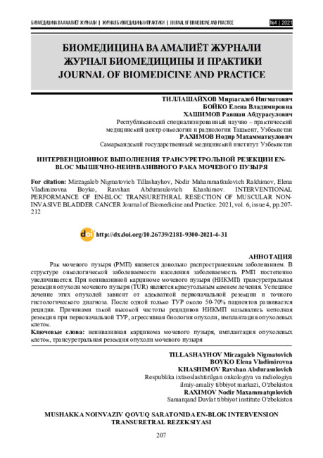 Interventional performance of en-bloc transurethral resection of muscular non-invasive bladder cancer
Journal of Biomedicine and PracticeBladder cancer (BC) is a rather common disease. The incidence of bladder cancer in the population is gradually increasing[3,7] In non-invasive bladder carcinoma (NICM), transurethral resection of bladder tumour (TUR) is the cornerstone of treatment. Successful treatment of these tumours depends on adequate initial resection and an accurate histological diagnosis. After TUR alone, around 50 70% of patients develop recurrence. Reasons for this high rate of recurrence of NICMP have been cited for incomplete resection during the initial TUR, aggressive tumour biology, and implantation of tumour cells[6,8,9].
-
 Results of modified surgical access to regional lymph nodes in the treatment of renal cancerPurpose: To study the long-term results of a new method of surgical access to the main vessels and regional renal cancer lymphocollectors. Materials and methods: In the Republican Specialized Scientific and Practical Medical Center of Oncology and Radiology, as well as in its Tashkent City Branch from 2010-2015, 96 patients were treated. The distribution of patients by sex and age intervals is shown in the figure. There male predominance 51 (53.1%) versus 45 (46.9%) women, the ratio was 1.1: 1. The age range is from 21 to 75 years, the average age of patients was 56.0 ± 1.3 years. From 96 patients participating in the study, a tumor process was established in 51 (53.1%) men and 45 (46.9%) women. The number of patients with metastatic lesion of regional lymph nodes were followings: N1 - 10 (10.5%), N2-6 (6.2%). In the remaining 80 (83.3%) patients, regional lymph nodes were intact. All patients underwent radical-extended nephrectomy. After laparotomy, the ascending and transversely dividing colon of the large intestine is mobilized, the loops of the intestines are pulled back with a moist towel up and to the left. After this, the back of the peritoneum is opened over the projection of the aorta from the level of the aortic bifurcation, the top with the dissection of the triceal ligament is opened beyond the peritoneal space. Results: The character of the histological form of the kidney tumors established after the operation by morphological examination were followings: the clear cell cancer in 77 (80.2%), papillary cancer in 8 (8.3%), unclassifiablc cancer in 4 (4.2% ), chromophobic cancer - in 7 (7.3%). At stage 1, no metastasis was detected. In stage 2 metastasis was noted in 4.2% of cases. In stage 3, 18.0% metastasis was detected. In 27.3% of patients metastasis took place in the 4th stage. All 13 patients (13.5%) had postoperative progression of the disease revealed. Conclusion: In renal tumors with Ti, we recommend performing selective lymphodisection, that is removing of first-order lymph nodes for precise staging. For T2.4N0.2M0 tumors, it is recommended to perform an extended lymph dissection from the level of the diaphragm legs to the aortic bifurcation. The development of surgical access with a cut of the triceal ligament with extended lymph node dissection of the renal cancer and a small number of postoperative complications make it possible to apply it in all patients with renal cancer.
Doctor's Herald -
 Kidney cancer regions limfatugunlarini-invasive visualization methodsIn order to check the Samarkand Regional Cancer Center, years 2002-2007, 101 patients with kidney cancer were controlled.They were treated in a specialized urology center of Samarkand Regional Cancer Center, Cancer Research Center. Among them, men 57, women -44. Male and female ratio of 1.27: 1. The cancer of right kidney had 56 patients, the cancer of left kidney-45 patients. The average age was 44.6 ± 1.4. Parakaval lymph node authenticated MRT sensitivity 90.3%, CT at 91%, the US accounted for 57.7%. CT specificity 68.6%, CT at 78.1% and MRT at -89.2%. Aortokoval lymph node identified changes in the MRT sensitivity of 92.0%, CT - 82.3%, CT - 68.0%. Specificity of ultrasound at 67.3%, CT - 84.4% and MRT was 93.4%. Paraaortic lymph node changed MRT sensitivity of 94.1%, CT - 82.3%, CT - 75%. An indication of the specificity of MRI at 95%, CT - 80% of the US accounted for 67.6%. Taking into account the above changes in renal cancer to the regional lymph nodes of non-invasive diagnostic method is recognized as the most effective method of MRT. As the MRT is better than SE, with its high sensitivity and specificity. Ac-cording to the ultrasound assessment, the changes in the regional lymph nodes may lead to the diagnostic error.
Journal problems of biology and medicine -
 IMPROVEMENT OF METHODS FOR EARLY DIAGNOSIS OF CERVICAL CANCER BASED ON LIQUID CYTOLOGY AND ONCOPROTEIN 53Cervical cancer remains a major preventive cancer (oncology) problem in both developed and developing countries, being the third leading cause of cancer mortality among women worldwide. Since the introduction of organized cytological screening programmes among the population, there has been a sharp decrease in the incidence of cervical cancer in many countries. Regardless of this encouraging result, the overall effectiveness of cervical cytologic screening of the cervix, the Papanicolaou smear is still far from optimal. Screening programmes using traditional cytology have successfully reduced the risk of cervical cancer, but new tests such as liquid cytology and human papillomavirus testing could improve screening and accuracy of the data received, and studying the p53 gene, which plays a key role in preventing tumors, could help treat malignancies. The purpose of this report was to study the descriptions of modem minimally invasive methods for determining cervical cancer risks and the possibility of using them in screening oroerammes.
Doctor's Herald -
Transurethral Resection Of En-Bloс Muscularis Non - Invasive Bladder Cancer
The American Journal of Medical Sciences and Pharmaceutical ResearchThe high recurrence rate of muscle noninvasive bladder cancer (BC) dictates the search for new methods of surgical treatment. The problem of bladder cancer (BC) treatment is very urgent in urology, because of high morbidity, difficulties in determining the optimal treatment tactics, necessity of long-term follow-up examinations, high recurrence rate and progression. Risk and progression criteria have been developed to identify groups of patients in need of closer follow-up, which can be quantified using risk calculators for recurrence and tumour progression. Although there are clear guidelines for the treatment of patients with bladder cancer, it is also believed that the rate of recurrence depends on the quality of the primary surgical procedure performed [1,2].
-
 Сравнительный анализ эффективности диагностических методов при опухолях носоглотки у детей и подростковЗлокачественные опухоли носоглотки у детей составляют 1-3% от всех злокачественных опухолей детского возраста и 10-12% от опухолей головы и шеи, а в структуре заболевания среди опухолевых заболеваний ЛОР-органов – составляют 25% [6, 9, 10].
Journal problems of biology and medicine -
 Ultrasound sonography in differential diagnosis of breast tumorsTo achieve the objectives 321 women were surveyed. Age of patients ranged from 24 to 82 years. Traditional ultrasound has enabled us to detect breast cancer in 96 observations, representing 83% of all pa-tients with breast cancer. Based on the analysis of ultrasound findings, it was concluded that the accuracy of conventional ultrasonography in differential diagnosis of palpable cancers is 84%, and 73% in difficult pal-pable cancers. The diagnostic accuracy of ultrasound examination was 84%. The main criteria for cancer with ultrasound are hypoechoic formation of irregular or indefinite shape, with indistinct contours and the vertical orientation of the long axis in the breast with respect to skin tissue. The diagnostic accuracy of ultra-sound in the diagnosis of cancer is difficult palpable does not exceed 73%. Verification of breast cancer, which is palpated, the procedure of fine-needle aspiration biopsy under ultrasound guidance is possible with an accuracy of 76%. The corresponding figure for the automatic trepanobiopsy - 91%. Verification of cancer is difficult palpable breast cancer procedure, fine needle aspiration biopsy under ultrasound guidance is pos-sible with an accuracy of 82%. The corresponding figure for the automatic trepanobiopsy - 88%
Journal problems of biology and medicine -
 Local staging of rectal cancer with conventional and diffusion-weighted magnetic resonance imagingIn this article we present up-to-date views on rectal cancer imaging. Accurate initial and follow-up staging of rectal cancer is vitally important and provides information essential for treatment decision making, preoperative therapy and surgery planning. Almost worldwide magnetic resonance imaging (MRI) is currently considered as the most advanced staging modality for rectal cancer; however conventional MRI sequences have some diagnostic limits and not always allow to differentiate fibrotic tissue (present either desmoplastic reaction or fibrotic changes due to preoperative radiotherapy) from tumor. On the basis of our own experience and published data we tried to demonstrate imaging possibilities of conventional and diffusion-weighted (DW) MRI in local staging of rectal cancer
Journal problems of biology and medicine -
 Cervical cancer prevention methods and human papilloma virus infectionIn the structure of malignant neoplasms in women in Uzbekistan, cervical cancer ranks second after breast cancer (BC), while its share is 12.49%. Standardized incidence rate CC in the republic in 2015 amounted to 5.5 per 100 thousand of the world population. The highest incidence of cervical cancer is characteristic at the age of 45-64 years, at the age of 0-19 years it practically does not occur (0.066%), the peak incidence occurs at 45-64 years, more than 28% of women fall ill before 44 years of age, while the average age falls on women 53 years old. Considering the statistical indicators of the incidence of cervical cancer, which tend to increase, the problem of studying this pathology from the point of view of oncoepidemiology, and also, the mechanisms of carcinogenesis is relevant and in demand. HPV is one of the determining factors contributing to the development of cervical cancer. Every year, 470 thousand new cases of cervical cancer are registered in the world, which is 14.2% of all malignant neoplasms in women, more than half of which ends in death. HPV is one of the studied and most common infections that predetermine the development of this pathology - papillomavirus, which is diagnosed in more than 3 million people. cases [1, 2, 3].
Journal problems of biology and medicine -
 The RSO and RSMC Samarkand included 68 patients with metacarpenic breast cancer from 2007 to 2017. More than half of the patients were women of reproductive age (26-49 years). The difference between the primary and metacarpal cancer in unilateral cancer was detected at a very early stage of the disease. This is because patients with cancer treatment are in deep dosing. Metaxron is one of the major factors affecting the survival of patients with ovarian cancer: the second stage of cancer, the size of the tumor in the lactation, the volume of treatment, the rate of damage to the regional lymph nodes and the patient's age
 -
 Comparative estimation of methods of treatment of a cancer of a bladder with metastasises in regional lympho noduses
Doctor's HeraldThe Tashkent Institute of improvement of doctors Results of treatment of 78 bladders sick by a cancer (BCa) with a lesion regional lymph nodes in stages T3-4 N1-2 MO are presented. The recurrent tumor is taped 19 (82,6 %) from 23 patients by whom the radical cystectomy (RCE) with standard lymph node dissection and the subsequent polychemotherapy (PCHT ) (I group was spent). On the average in 5,2 months after the treatment termination; in 11 group of 25 patients after RCE with dilated lymph node
dissection and the subsequent PCHT at 18 (72.0 %) the recurrent tumor, on the average in 4,8 months is taped. In 111 group after carrying out PCHT (without surgical treatment) advance of growth of a tumor after its regress and stabilization, was diagnosed at 27 (90,0 %) from 30
patients, on the average in 3,5 months. The survival rate median in 1 group of patients has made 9,4± 3,6 months, in II group - 12,5±4,2 months and in 111 group - 7,4 ± 2.9 months (p <0,05) -
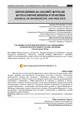 Cervical cancer during pregnancy (modern aspects of diagnostics and tactics of management). (Literature review)
Journal of Biomedicine and PracticeDiagnosis and management of pregnant women with cervical cancer is a difficult problem for clinicians. There is still no consensus on the need for a biopsy or conization in pregnant women, especially with suspected CIN III and cancer in situ. The issues of diagnosis, treatment, delivery and monitoring in pregnant women with cervical cancer are practically not covered in textbooks and scientific and practical publications. This article analyzes the recommendations of the international scientific community and the results of large-scale clinical studies on the management of pregnant women with abnormal cytological smears during pregnancy. Summarized current knowledge about the diagnosis and treatment of CIN during pregnancy. Also reviewed and analyzed more than 40 works of domestic and foreign authors on this issue
-
 The incidence of kidney cancer in Samarkand region. the present state of diagnostics and treatmentThe article retrospectively analyzed the incidence of kidney cancer in the Samarkand region in the peri-od from 2006 to 2017, and the status of diagnostics and treatment. In the structure of cancer pathology in Sa-markand region kidney cancer constitutes 2.7% and takes 12th place. We may observe increase in the inci-dence of kidney cancer over the last 7 years up to 1.8 times and a low detection of process in early stages. This study is based on the diagnostics and treatment results of 415 patients with kidney cancer. Diagnostic status until 2015 in the area did not meet the requirements of the standard. Over the past 3 years in connection with the introduction of the technique needle biopsy, percutaneous nephrostomy and appearance of ability to apply MSCT, dramatically increased the efficiency of diagnosis, and that influenced the treatment results. Radical nephrectomy performed 162 (39,0%) patients, palliative 63 (15,2%) patients. Chemo - and immunotherapy 53 (12,8%), immunotherapy 62 (14.9 %) and more modern targeted therapy 12 (2,9%) patients, 58 (14,0%) pa-tients underwent symptomatic therapy and in 5 (1,2 %) patients treatment was not conducted
Journal problems of biology and medicine -
Analysis Of Primary Oncological Disability In Andijan Region In 2017-2018
The American Journal of Medical Sciences and Pharmaceutical ResearchMalignant neoplasms are one of the most relevant areas of modern medicine. More than 120 thousand people annually. recognized as disabled due to oncological diseases. In the structure of mortality from this class of diseases, about 30% are people of working age [1,5,9].
In the structure of primary disability, malignant neoplasms occupy the 2nd place after diseases of the circulatory system (16-20% of the total number of those who were first recognized as disabled) [1,2,9].
Affected mainly by the population in active working age, up to 90-95% of patients at the initial examination are recognized as invalids of groups I-II [22].
In men, the main disabling disease is lung cancer - 27.8%, in second place - stomach cancer - 11.3%, in third - cancer of the rectum and colon - 11.0%, in fourth - cancer of the larynx - 7.3%. In women, the leading disabling disease is breast cancer - 39.5% and female genital cancer - 26.3%. Cancer of the rectum and colon occupies 9.2% in the structure of primary disability in women [11,12,18].
-
ILLUMINATING CANCER DETECTION: SPECTROSCOPY'S VITAL ROLE IN SCREENING AND DIAGNOSIS
The American Journal of Applied sciencesThis paper explores the pivotal role of spectroscopy in cancer screening and diagnosis, shedding light on its significance in early detection and accurate characterization of cancerous tissues. Spectroscopic techniques offer unique capabilities for non-invasive and real-time analysis of tissue composition, providing valuable insights into biochemical and structural alterations associated with cancer development. By leveraging the inherent molecular signatures of tissues, spectroscopy enables clinicians to identify abnormal changes indicative of cancer presence, facilitating timely intervention and improved patient outcomes. This review examines the principles, methodologies, and applications of spectroscopy in cancer detection across various modalities, including Raman spectroscopy, fluorescence spectroscopy, and infrared spectroscopy. Additionally, it discusses recent advancements, challenges, and future directions in harnessing spectroscopic technologies for enhanced cancer screening and diagnosis.
-
 Результаты лечения больных раком прямой кишки при использовании различных методов предоперационной лучевой терапииНаше исследование основано на клинических наблюдениях над 106 больными раком прямой кишки с T3N0-2M0 стадией заболевания, которые получали комбинированное лечение в условиях РОНЦ с 2002 по 2006 гг.
Journal problems of biology and medicine